Femoroacetabular joint
The femoral head fits deeply into the acetabulum, providing substantial bony stability while still allowing flexion, extension, rotation, and abduction.
Hip · The hip is a deep, stable ball-and-socket joint that bears several times body weight during walking and running, making it a common site of both overuse pain and degenerative change.
Darker cells mean a higher score for this topic on that metric.
LessMore
Last reviewed Sources & creditsMedia creditsMethodology
Osteoarthritis is among the most common causes of hip pain after middle age, involving gradual breakdown of the joint's cartilage and associated changes in the surrounding bone and soft tissue.
Greater trochanteric pain syndrome describes pain at the outer hip related to irritation of the gluteal tendons or the nearby bursa, often mistaken in the past for bursitis alone.
Femoroacetabular impingement describes abnormal contact between the femoral head and the hip socket during certain movements, related to subtle variations in bone shape, which can contribute to groin pain and reduced range of motion in some individuals.
Many cases of hip osteoarthritis are managed for years with exercise, weight management, and activity modification; joint replacement is typically considered when conservative measures no longer provide adequate relief.
Reduced strength or endurance in the gluteal muscles is commonly associated with several hip and even knee conditions, since these muscles are central to controlling pelvis and leg alignment during walking and running.
A hip labral tear is damage to the ring of cartilage that deepens the hip socket, which can occur from trauma, repetitive impingement, or gradual wear, and may contribute to pain, clicking, or a sense of instability.
The hip joint is formed where the rounded head of the femur fits into the deep, cup-shaped acetabulum of the pelvis, an arrangement that provides substantial inherent stability compared with more shallow joints like the shoulder.
A large group of muscles surrounds the joint, including powerful hip flexors at the front, extensors at the back, and abductors at the side that are essential for controlling pelvic stability during single-leg activities such as walking and running.
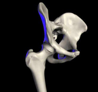
The femoral head fits deeply into the acetabulum, providing substantial bony stability while still allowing flexion, extension, rotation, and abduction.
A ring of cartilage around the rim of the socket increases joint contact area and stability, and can be a source of pain when torn.
This hip abductor muscle controls pelvic tilt during walking and running, and its weakness is commonly associated with greater trochanteric pain syndrome and altered gait mechanics.
This deep muscle group flexes the hip and can become a source of anterior hip or groin pain when irritated, particularly in activities involving repetitive hip flexion such as kicking or sprinting.
The hamstring muscles originate at the base of the pelvis and extend the hip, and this attachment point can become a source of deep buttock pain when irritated.
A thick fibrous capsule reinforced by several ligaments limits excessive joint motion and works alongside the surrounding muscles to maintain stability.
Hip flexion, bringing the knee toward the chest, represents the joint's largest range of motion, while extension is considerably more limited at roughly 10–20° due to the tension of anterior hip structures.
This combination of deep bony stability and powerful surrounding muscles explains why hip pain can originate from the joint itself, the surrounding tendons, or the muscles that control pelvic movement, each requiring a different assessment focus.
Neighbouring topics share region literacy — not a severity ranking.
The knee is the body's largest joint and one of its most frequently injured, relying on ligaments and surrounding muscles rather than bony shape for stability.
Pain frequency 65 Same body regionThe anterior cruciate ligament stabilizes the knee against pivoting and forward-sliding forces, and its rupture is one of the most studied and rehabilitation-intensive sports injuries.
Pain frequency 50 Same body regionThe ankle and foot absorb and redirect the full force of body weight with every step, making sprains and overuse injuries among the most common in sport and daily life.
Pain frequency 70 Same body region