18 min read
If you only read one thing
Electrocorticography and depth electrodes — millimetre-scale human recordings that reveal high-frequency dynamics surgery can see.
Key takeaways
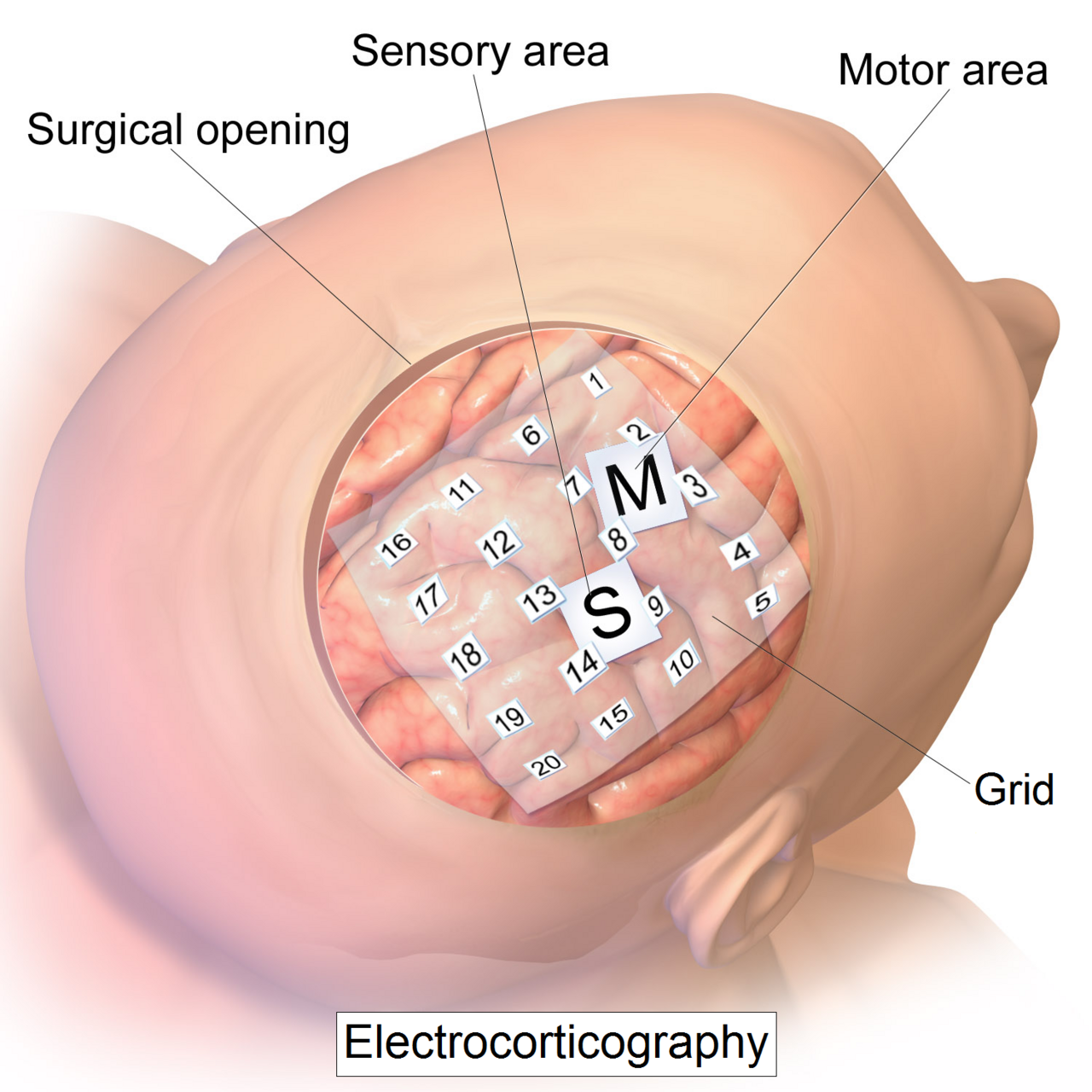
Intracranial EEG records electrical activity from electrodes on the cortical surface (ECoG) or in depth (stereo-EEG / SEEG). It offers millimetre spatial scale and kilohertz temporal bandwidth unavailable to scalp EEG or fMRI. Clinically it maps seizure onset and eloquent cortex before epilepsy surgery; scientifically it reveals high-gamma activity tied to cognition. Coverage is sparse and ethical constraints are strict — not a general “brain reading” tool. Educational neuroscience context only — not medical advice. Intracranial EEG is performed in specialised clinical settings for indicated patients.
Jump to section
Previous
PTSD
Next
Brenda Milner
Who this is for Electrocorticography and depth electrodes — millimetre-scale human recordings that reveal high-frequency dynamics surgery can see.
Print this page
Key scores Research 82 Awareness 45 Complexity 85 Clinical weight 75 Plasticity 40 Live debate 60 Memory Attention Language Emotion
Key takeaways
Focus: invasive signals Research: 82/100 Awareness: 45/100 Complexity: 85/100
Open brain atlas →
Quick facts invasive signals Focus
82/100 Research
45/100 Awareness
85/100 Complexity
75/100 Clinical
60/100 Debate
Intracranial EEG records electrical activity from electrodes on the cortical surface (ECoG) or in depth (stereo-EEG / SEEG). It offers millimetre spatial scale and kilohertz temporal bandwidth unavailable to scalp EEG or fMRI.
Clinically it maps seizure onset and eloquent cortex before epilepsy surgery; scientifically it reveals high-gamma activity tied to cognition. Coverage is sparse and ethical constraints are strict — not a general “brain reading” tool.
Educational neuroscience context only — not medical advice. Intracranial EEG is performed in specialised clinical settings for indicated patients.
Local lens clinics, schools and research labs.
Educational atlas — not a diagnosis, treatment plan, or medical advice.
Methods.
Start here mm ECoG millimetre timing
82 Research editorial band
1950s Penfield awake mapping teaching history
0 OR recipe no OR checklist here
Orient — Intracranial EEG — MethodsCompare — Use compare / related rails for neighbours.Go deep — AnatomyCheck local — Check localLens before treating English norms as universal.Desk note Primary desk: clinics, schools and research labs.
Note Educational atlas — not a diagnosis, treatment plan, or medical advice.
Limit clinics, schools and research labs
Scope Editorial scores are browsing aids, not certificates.
Primary — clinics, schools and research labsSecondary — MethodsMethod — Editorial synthesis — prefer primary desks for decisions.Intracranial EEG — concrete atlas depth on Tool-Lifes.
Local source desk Clinic vs school support here Care pathways and school supports are national. This atlas is educational — not a diagnosis.
Clinic door GP / neurology / memory clinic (NHS or insurance referral)
School support IEP / 504-style plans — names differ; ask the school, not a blog
Research vs care Lab findings are not a treatment plan
Named sources NINDS / NICE-style public pages — dated editions Keep this desk beside the atlas when a school or clinic conversation is open.
Not a diagnosis, treatment plan or medical advice. Scores are browsing aids.
Methods Methods — clinics, schools and research labs.
Limit Educational atlas — not a diagnosis, treatment plan, or medical advice.
Field terms
Local lens Everyday desks for this language community.
Editorial score 0–100 browsing aid inside this atlas only.
Caveat Educational atlas — not a diagnosis, treatment plan, or medical advice.
Primary desk clinics, schools and research labs
Start path overview → anatomy → function
FAQ Eight distinct questions per topic page.
Share blurb Native one-liner for social previews.
Best-30 Quality pass for locale, traffic and premium rails. How to use this atlas Start here — OverviewNext — AnatomyThen — FunctionCheck local — Local lensTools Open the brain quiz
Local systems
English-speaking clinics and labs DSM/ICD teaching frames - not a diagnosis
hospital neurology and psychology labs schools, clinics and research centres
overview methods · Intracranial EEG Local examples schools, clinics and research centres hospital neurology and psychology labs Prefer local desks before English defaults. Quick FAQ What is intracranial EEG iEEG/ECoG? Electrodes on or in the brain during clinical mapping — millimetre-scale timing that scalp EEG blurs. Teaching description of published methods.
When did Penfield-era cortical stimulation map speech? Mid-20th-century Montreal work made awake mapping famous. Modern grids and stereo-EEG continue the classroom story without copying an OR checklist.
Is this a guide to implant surgery or dosing? No. Educational methods atlas. Not a diagnosis, implant protocol, or medication dose.
Related searches Intracranial EEG What is intracranial Intracranial EEG atlas Intracranial EEG What can Intracranial EEG numbers
How to read this page Start here — Overview — Intracranial EEGNext — Anatomy · clinicsThen — Function · MethodsIntracranial EEG — concrete atlas depth on Tool-Lifes.
Editorial timeline 1 — overview · Intracranial EEG2 — anatomy · hospital neurology and psychology labs3 — function · methods2026 — Editorial review stampPrimary desks Primary frame — DSM/ICD teaching frames - not a diagnosisSecondary frame — hospital neurology and psychology labsMethod note — schools, clinics and research centresNote Educational atlas — not a diagnosis, treatment plan, or medical advice.
The profile
82 45 85 75 40 60
Research 82 Awareness 45 Complexity 85 Clinical weight 75 Plasticity 40 Live debate 60 Seven ways into this topic Frequently asked questions What is intracranial EEG iEEG/ECoG? Electrodes on or in the brain during clinical mapping — millimetre-scale timing that scalp EEG blurs. Teaching description of published methods.
When did Penfield-era cortical stimulation map speech? Mid-20th-century Montreal work made awake mapping famous. Modern grids and stereo-EEG continue the classroom story without copying an OR checklist.
Is this a guide to implant surgery or dosing? No. Educational methods atlas. Not a diagnosis, implant protocol, or medication dose.
What can iEEG resolve that scalp methods smear? When and where populations engage during perception, speech, memory or movement — at scales scalp EEG averages away.
Research score 82 — a clinic ranking? No. Editorial 0-100 in this methods field. Compare with EEG, fMRI and MEG neighbours.
Why do overview pages get clicks? They name ECoG sites and cortical surface mapping early. Keep reading Overview, Anatomy, Function; do not self-schedule a procedure.
What reusable tool sits on brain pages? Atlas dual-view: pin a cortical label beside a deep structure while you read iEEG limits — teaching, not targeting.
Any number to keep? Scores are editorial. Clinical sampling density is case-specific in the literature — this site does not publish an OR recipe.
Previous
Diffusion MRI