Primary joint surfaces
Load-bearing surfaces of the neck region that guide available motion and contact stress.
Neck · The seven cervical vertebrae and the soft tissues that carry the head — posture load, disc and facet pain, and graded mobility work.
Darker cells mean a higher score for this topic on that metric.
LessMore
Last reviewed Sources & creditsMedia creditsMethodology
Not always. Sensitivity can rise with load spikes, sleep loss and stress even when imaging is unremarkable. Clinicians separate serious pathology from common nociplastic or mechanical patterns.
Many soft-tissue presentations settle over weeks to a few months with graded loading; post-operative and neuro pathways often run longer and are criteria-based.
Guidelines generally reserve early imaging for red flags or when results would change management — not for every first presentation of mechanical pain.
Transient soreness after new loading is common; sharp, escalating, night-waking or neurological change is a reason to stop and reassess with a clinician.
They can be adjuncts for confidence or tissue protection in defined windows, but durable capacity usually comes from progressive loading and skill practice.
Red flags, progressive neurological deficit, failed adequate conservative care, or surgical candidacy discussions — timing varies by condition and health system.
Understanding neck anatomy clarifies why certain loads provoke symptoms and why neighbouring joints are often trained together.
Rehab language mixes osteology, peri-articular soft tissue and sensorimotor control — all three appear in well-designed programmes.
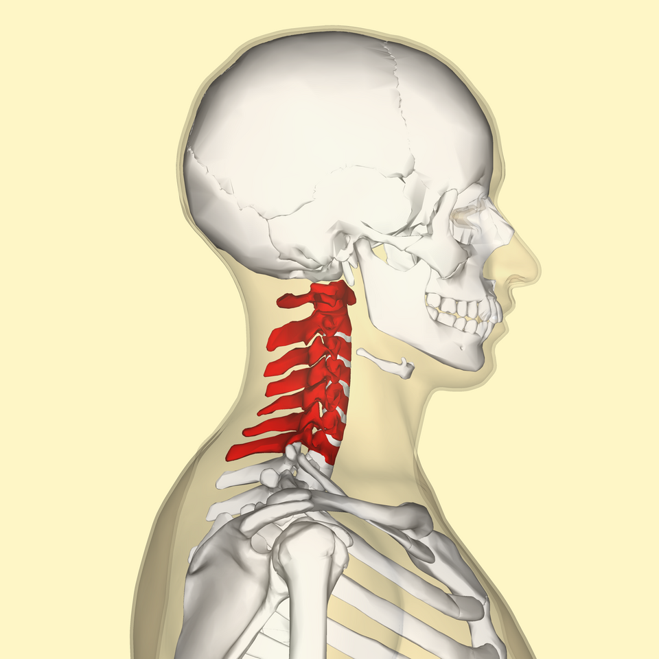
Load-bearing surfaces of the neck region that guide available motion and contact stress.
Ligamentous checks that set end-range and protect against excessive translation or rotation.
Larger muscles that produce the primary moments used in daily tasks and sport.
Shorter, often deeper fibres that fine-tune segmental stiffness and joint centring.
Peripheral nerves and autonomic supply that can refer symptoms or limit tolerance when irritated.
Bursae and fascial planes that allow neighbouring tissues to slide under repetitive motion.
Published goniometric norms for neck vary by age and method; clinicians compare side-to-side and task demands rather than chasing a single textbook number.
Anatomy pages here are maps for literacy, not a substitute for hands-on assessment of an individual joint.
Neighbouring topics share region literacy — not a severity ranking.
The lumbar spine bears more compressive load than any other spinal region, making low back pain the single most common musculoskeletal complaint worldwide.
Pain frequency 80 Same body regionThe core is the coordinated group of trunk muscles that stabilizes the spine and pelvis, transferring force between the upper and lower body during nearly every movement.
Pain frequency 35 Same body region